JEEP
YJ-SERIES Catalogue
Fiberglass
and Steel Parts
If a part begins with
the letter "G" under "PART#" it is referring
to fiberglass.
Any other instances are describing steel parts. Much more coming
soon!!
A picture gallery is included along with full price list.
See below table for gallery.
Thanks for your
interest. Please call
or Email any needs. Contact the company
by e-mailing (click
link) or telephone (905) 857-6345.
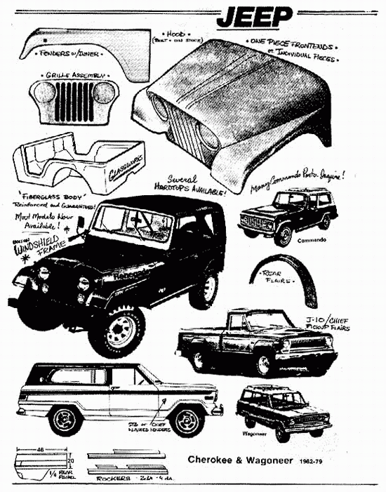
PICTURES BELOW-LOTS MORE COMING SOON.........PLEASE STAY TUNED!!!!!!!!!!!
JEEP YJ / WRANGLER 87-96 REPRODUCTION STEEL
Keep event rate under 1,000-2,000 events/second. High speed distorts FSC-A due to pulse overlap.
Use FSC-A vs. FSC-H (or FSC-A vs. FSC-W) to remove doublets before analyzing DNA content. The purity of your G1 and G2 peaks depends entirely on this gate. 2. Viability and Apoptosis Assays Dead cells have lower FSC-A than live cells (they shrink and lose membrane integrity). However, debris also has low FSC-A. By combining FSC-A with SSC-A (Side Scatter – Area), you can cleanly separate live cells from debris. Be cautious: highly apoptotic cells can fragment, and those fragments will have very low FSC-A. 3. Immunophenotyping (Leukocyte gating) In whole blood or spleen analysis, FSC-A vs. SSC-A is the classic first gate. Lymphocytes (low FSC-A, low SSC-A), monocytes (high FSC-A, low SSC-A), and granulocytes (high FSC-A, high SSC-A) form distinct populations. Remember that FSC-A here is relative—activation of lymphocytes (e.g., blast formation) increases FSC-A, while red blood cell lysis artifacts can decrease it. 4. Cell Sorting (FACS) When sorting cells, the sorter uses FSC-A to decide when to charge a droplet. However, doublets confuse sorters. By strictly gating on the FSC-A/FSC-H diagonal, you ensure that you are sorting true single cells, preventing clogged nozzles and improving post-sort viability. Part 4: Troubleshooting Common FSC-A Problems Problem 1: "My FSC-A signal is off scale" Symptoms: A flat line at the top of the plot; populations look "squished." Cause: Gain is too high. Solution: Use beads (e.g., 3µm and 6µm) to set voltages. For most cells (3-15µm), start with FSC voltage at ~50-100V on analyzers (e.g., BD LSRFortessa). Never use automatic FSC gain on unknown samples – it will ruin relative size comparisons. Problem 2: Poor separation of live vs. dead Symptoms: No distinct population; debris overlapping with live cells. Cause: FSC-A alone is insufficient. Solution: Use a viability dye (e.g., 7-AAD, PI, or fixable live/dead stains). FSC-A is a physical parameter; viability dyes are chemical . The combination is powerful. Problem 3: Doublets are not obvious on FSC-A vs. FSC-H Symptoms: A diagonal line with no clear off-diagonal population. Cause: Your sample is mostly single cells, OR your flow rate is too high. High event rates (>5,000 events/sec) cause coincidence (two cells passing simultaneously but not adhered), which can mimic singlet behavior. Solution: Reduce flow rate to <2,000 events/sec and re-analyze. Problem 4: Comparing FSC-A across experiments Reality: You cannot reliably compare absolute FSC-A values between different days or different instruments unless you use standardized beads (e.g., Cytometer Setup and Tracking beads). Even then, FSC is highly sensitive to laser alignment, fluidics, and temperature. For quantitative size comparisons, use calibrated beads (e.g., SpheroTech) to convert FSC-A into microns. Part 5: Step-by-Step Optimization Protocol for FSC-A If you are setting up an experiment today, follow this protocol: Keep event rate under 1,000-2,000 events/second
To exclude doublets, gate only the cells where FSC-A ≈ FSC-H (the diagonal). Part 3: Practical Applications – Where FSC-A Shines 1. Cell Cycle Analysis (Propidium Iodide / DAPI) This is the most common application where FSC-A is non-negotiable. In DNA content analysis, doublets are disastrous because a doublet of G1 cells (2N each) will mistakenly appear as a single G2/M cell (4N DNA). This ruins your cell cycle modeling. FSC-H (or FSC-A vs
In your methods section, always report: "Doublets were excluded using FSC-A/FSC-H singlet gating." Part 6: Advanced Considerations and Variants Cytometers Without FSC-A (e.g., some benchtop models) Older or simpler cytometers (like the first-generation Guava systems or some CytoFLEX configurations) may not report FSC-H or FSC-W. In these cases, you cannot perform traditional doublet discrimination. Alternatives include using SSC-A vs. SSC-H or fluorescence pulse geometry (e.g., PI-A vs. PI-W in cell cycle). Spectral Flow Cytometry In spectral cytometers (e.g., Cytek Aurora), the concept of FSC-A remains, but the traditional photodiode is replaced. However, the physics of forward scatter is unchanged. Crucially, spectral cytometers often allow unmixing of scatter parameters, but FSC-A remains a vital doublet discrimination tool. Imaging Flow Cytometry (e.g., Amnis ImageStream) Here, "FSC-A" is calculated from the image mask. While less common, the same principle applies: area vs. height (or aspect ratio) weeds out doublets and clusters. However, imaging provides the ultimate confirmation – you can literally see if it’s a doublet. Conclusion: Why FSC-A Deserves Your Respect In the rush to analyze bright fluorescent markers, many researchers treat FSC-A as an afterthought—an "auto" setting they click and forget. This is a mistake. Poor FSC-A gating leads to doublet contamination, skewed cell counts, and irreproducible results. Good FSC-A gating, conversely, is the hallmark of a rigorous flow cytometrist. skewed cell counts
Run a mix of small (3µm) and large (6-10µm) beads to check the dynamic range. Adjust FSC voltage so both populations are on scale (usually between 10^2 and 10^5 on a log scale or 100-200K on a linear scale).